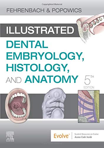
Get a clear picture of oral biology and the formation and study of dental structures. Illustrated Dental Embryology, Histology, & Anatomy, 5th Edition is the ideal introduction to one of the most foundational areas in the dental professions – understanding the development, cellular makeup, and physical anatomy of the head and neck regions. Written in a clear, reader-friendly style, this text makes it easy for you to understand both basic science and clinical applications – putting the content into the context of everyday dental practice. New for the fifth edition is evidence-based research on the dental placode, nerve core region, bleeding difficulties, silver diamine fluoride, and primary dentition occlusion. Plus, high-quality color renderings and clinical histographs and photomicrographs throughout the book, truly brings the material to life.
- UPDATED!
Test Bank with cognitive leveling
- and mapping to the dental assisting and dental hygiene test blueprints.
- UPDATED! User-friendly pronunciation guide of terms ensures you learn the correct way to pronounce dental terminology.
- Comprehensive coverage includes all the content needed for an introduction to the developmental, histological, and anatomical foundations of oral health.
- Hundreds of full-color anatomical illustrations and clinical and microscopic photographs accompany text descriptions of anatomy and biology.
- Clinical Considerations boxes relate abstract-seeming biological concepts to everyday clinical practice.
- Key terms open each chapter, accompanied by phonetic pronunciations, and are highlighted within the text, and ag glossary provides a quick and handy review and research tool.
- Expert authors provide guidance and expertise related to advanced dental content.
- NEW!
Evidence-based research
- thoroughly discusses the dental placode, nerve core region, bleeding difficulties, silver diamine fluoride, and primary dentition occlusion.
- NEW! Photomicrographs, histographs, and full-color illustrations throughout text helps bring the material to life.
- NEW! The latest periodontal insights include biologic width, gingival biotype, gingival crevicular fluid quantitative proteomics, clinical attachment level, AAP disease classification, and reactive oxygen species therapy.
- NEW! Expanded coverage of key topics includes figures on tongue formation, developmental disturbances, root morphology, and TMJ cone beam CT.
Product Details
- Paperback: 352 pages
- Publisher: Saunders; 5 edition (December 2, 2019)
- Language: English
- ISBN-10: 0323611079
- ISBN-13: 978-0323611077
- Product Dimensions: 8.2 x 0.5 x 10.5 inches

