About This CME Teaching Activity
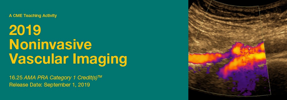
This CME activity offers a comprehensive review of recent developments and issues in noninvasive vascular imaging. Throughout the program faculty address state-of-the-art ultrasound and Doppler techniques and how they are used to evaluate the cerebrovascular, abdominal and peripheral vascular systems. In addition, diagnostic pearls and pitfalls, credentialing requirements, venous testing and the clinical applications of transcranial Doppler are discussed.
Target Audience
This CME Activity is intended and designed to offer educational opportunities to radiologists, vascular surgeons, cardiologists, sonographers, and other clinicians interested in learning more about noninvasive vascular ultrasound.
Scientific Sponsor
Educational Symposia
Educational Objectives
At the completion of this CME activity, subscribers should be able to:
- Discuss the newest techniques in the noninvasive diagnosis of endovascular disease, including venous and carotid imaging.
- Assess new developments in vascular imaging.
- Discuss the utility of ultrasound and Doppler imaging when used to evaluate the aorta, carotid arteries, and peripheral vascular system.
- Utilize ultrasound to evaluate abdominal transplants, endostents, and dialysis fistula.
- Utilize ultrasound to evaluate the venous system of the upper and lower extremities.
- Describe the appropriate role of ultrasound in the evaluation of the renal and hepatic arteries.
No special educational preparation is required for this CME activity
Topics/Speaker
1. Fundamentals The Carotid Duplex Exam
2. Evolution of Carotid Diagnostic Criteria
3. Update on IAC Carotid Diagnostic Criteria Project
4. Assessment of Vertebral Arteries and Arch Vessels in the Vascular Lab
5. Non Atherosclerotic Carotid Disorders
6. Fundamentals of the TCD Examination
7. Panel Our Most Interesting Carotid Cases of the Year
8. Fundamentals Arterial Duplex and Physiological Testing for Native and Revascularized Arteries
9. Doppler Waveform Nomenclature From Chaos to Standardization
10. Management of PAD New Developments in Medical Therapy
11. Guidelines for Follow-up after Arterial Procedures
12. Evaluation of Upper Extremity Arterial Disorders in the Vascular Lab
13. Our Most Interesting Arterial Cases of the Year
14. Too Many Images Over Documentation in the Vascular Lab
15. Lab Management Panel Pearls of Wisdom on Running a Vascular Lab
16. Screening and Follow-up of AAA – A Guideline Based Approach
17. Endoleak Detection after EVAR
18. Duplex Criteria for Native and Stented Mesenteric and Renal Arteries
19. 5 Top Tips on Renal Artery Imaging
20. Panel Our Most Interesting Abdominal Duplex Cases of the Year
21. Duplex Evaluation of Pelvic Venous Disorders
22. Advanced TCD Applications
23. Access Site Complications and Their Management
24. Hepatoportal Doppler and Evaluation of Liver Transplants
25. Fundamentals Venous Duplex Exam for Diagnosis of Lower and Upper Extremity DVT
26. Guideline Based DVT Management in 2019 – What the Vascular Lab Community Should Know
27. Non-DVT Must Knows for a Reader of Venous Duplex Studies Leslie M. Scoutt, M.D
28. Venous Reflux Testing Protocol, Technical Tips and Tricks
29. Assessment of Hemodialysis Access in the Vascular Lab
30. Mapping Prior to Dialysis Access Creation
31. Panel Our Most Interesting Venous Cases of the Year
32. Introduction to Contrast Enhanced Ultrasound
33. Contrast Enhanced Ultrasound of the Liver
34. Contrast Enhanced Ultrasound of the Kidneys
35. Vascular Emergencies
36. Carotid US 2019 Update
37. A Potpourri of Interesting Cases

